CD4 molecule
CD4分子 |
|---|

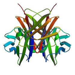
Crystallographic structure of the V-set and C2 domains of human CD4.[1] |
| 有效结构 |
|---|
| PDB | 直系同源检索:PDBe, RCSB |
|---|
| PDB查询代码列表 |
|---|
1CDH, 1CDI, 1CDJ, 1CDU, 1CDY, 1G9M, 1G9N, 1GC1, 1JL4, 1Q68, 1RZJ, 1RZK, 1WBR, 1WIO, 1WIP, 1WIQ, 2B4C, 2JKR, 2JKT, 2KLU, 2NXY, 2NXZ, 2NY0, 2NY1, 2NY2, 2NY3, 2NY4, 2NY5, 2NY6, 2QAD, 3B71, 3CD4, 3JWD, 3JWO, 3LQA, 3O2D, 3S4S, 3S5L, 3T0E, 4JM2 |
|
|
| 标识 |
|---|
| 代号 | CD4; CD4mut |
|---|
| 扩展标识 | 遗传学:186940 鼠基因:88335 同源基因:513 ChEMBL: 2754 GeneCards: CD4 Gene |
|---|
|
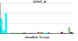
| RNA表达模式 |
|---|
 |
| 更多表达数据 |
| 直系同源体 |
|---|
| 物种 | 人类 | 鼠类 | |
|---|
| Entrez | 920 | 12504 | |
|---|
| Ensembl | ENSG00000010610 | ENSMUSG00000023274 | |
|---|
| UniProt | P01730 | P06332 | |
|---|
| mRNA序列 | NM_000616 | NM_013488 | |
|---|
| 蛋白序列 | NP_000607 | NP_038516 | |
|---|
| 基因位置 | Chr 12:
6.9 – 6.93 Mb | Chr 6:
124.86 – 124.89 Mb | |
|---|
| PubMed查询 | [1] | [2] | |
|---|
|
| CD4受体 |
|---|
 |
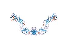
| structure of t-cell surface glycoprotein cd4, monoclinic crystal form |
| 鉴定 |
|---|
| 标志 | CD4-extrcel |
|---|
| Pfam(蛋白家族查询站) | PF09191 |
|---|
| InterPro(蛋白数据整合站) | IPR015274 |
|---|
| SCOP(蛋白结构分类数据站) | 1cid |
|---|
| OPM家族(膜蛋白方向) | 230 |
|---|
| OPM蛋白(膜蛋白方向) | 2klu |
|---|
| CDD(保守域数据站) | cd07695 |
|---|
|
CD4受体,全称“表面抗原分化簇4受体”(Cluster of Differentiation 4 receptors),是辅助T细胞的表面标记(surface markers)之一,也是辅助T细胞行使其功能的重要受体。当抗原呈递细胞(主要是巨噬细胞、棘状细胞及B细胞本身)将外来病菌分解,把抗原与主要组织相容性复合体结合后,呈递给辅助T细胞(即与辅助T细胞表面的CD4受体结合),辅助T细胞再接着刺激B细胞产生抗体,此即体液性免疫反应的基本过程。
资料
常用单克隆抗体或代号:T4,Leu3a 主要表达细胞:Tsub,Msub,Thysub T细胞 分子质量(kDa)和结构:gp55(IgSF) 功 能:与MCHn类分子结合,信号转导,HIV受体
参见
参考文献
- ↑ PDB 1cdh; Ryu SE, Truneh A, Sweet RW, Hendrickson WA. Structures of an HIV and MHC binding fragment from human CD4 as refined in two crystal lattices. Structure. January 1994, 2 (1): 59–74. PMID 8075984.
延伸阅读
- Miceli MC, Parnes JR. Role of CD4 and CD8 in T cell activation and differentiation. Adv. Immunol.. Advances in Immunology. 1993, 53: 59–122. doi:10.1016/S0065-2776(08)60498-8. ISBN 978-0-12-022453-1. PMID 8512039.
- Geyer M, Fackler OT, Peterlin BM. [http//www.ncbi.nlm.nih.gov/pmc/articles/PMC1083955/ Structure–function relationships in HIV-1 Nef]. EMBO Rep.. 2001, 2 (7): 580–5. doi:10.1093/embo-reports/kve141. PMID 11463741. PMC 1083955.
- Greenway AL, Holloway G, McPhee DA, et al.. HIV-1 Nef control of cell signalling molecules: multiple strategies to promote virus replication. J. Biosci.. 2004, 28 (3): 323–35. doi:10.1007/BF02970151. PMID 12734410.
- Bénichou S, Benmerah A. [The HIV nef and the Kaposi-sarcoma-associated virus K3/K5 proteins: "parasites"of the endocytosis pathway]. Med Sci (Paris). 2003, 19 (1): 100–6. doi:10.1051/medsci/2003191100. PMID 12836198.
- Leavitt SA, SchOn A, Klein JC, et al.. Interactions of HIV-1 proteins gp120 and Nef with cellular partners define a novel allosteric paradigm. Curr. Protein Pept. Sci.. 2004, 5 (1): 1–8. doi:10.2174/1389203043486955. PMID 14965316.
- Tolstrup M, Ostergaard L, Laursen AL, et al.. HIV/SIV escape from immune surveillance: focus on Nef. Curr. HIV Res.. 2004, 2 (2): 141–51. doi:10.2174/1570162043484924. PMID 15078178.
- Hout DR, Mulcahy ER, Pacyniak E, et al.. Vpu: a multifunctional protein that enhances the pathogenesis of human immunodeficiency virus type 1. Curr. HIV Res.. 2005, 2 (3): 255–70. doi:10.2174/1570162043351246. PMID 15279589.
- Joseph AM, Kumar M, Mitra D. Nef: "necessary and enforcing factor" in HIV infection. Curr. HIV Res.. 2005, 3 (1): 87–94. doi:10.2174/1570162052773013. PMID 15638726.
- Anderson JL, Hope TJ. HIV accessory proteins and surviving the host cell. Current HIV/AIDS reports. 2005, 1 (1): 47–53. doi:10.1007/s11904-004-0007-x. PMID 16091223.
- Li L, Li HS, Pa, et al.. Roles of HIV-1 auxiliary proteins in viral pathogenesis and host-pathogen interactions. Cell Res.. 2006, 15 (11–12): 923–34. doi:10.1038/sj.cr.7290370. PMID 16354571.
- Stove V, Verhasselt B. Modelling thymic HIV-1 Nef effects. Curr. HIV Res.. 2006, 4 (1): 57–64. doi:10.2174/157016206775197583. PMID 16454711.
外部链接
参考来源


